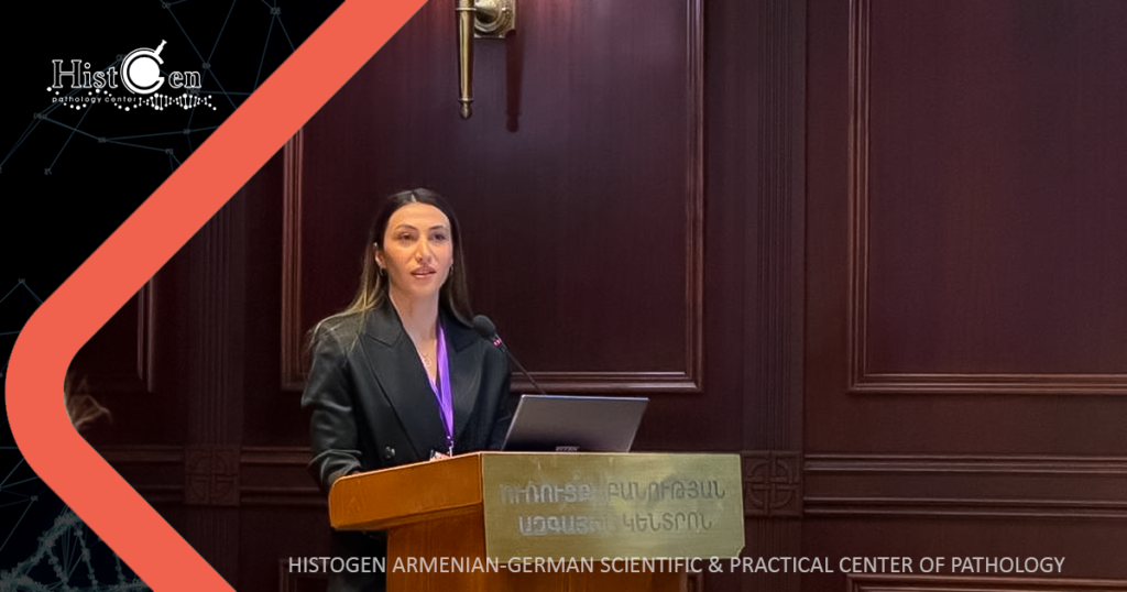
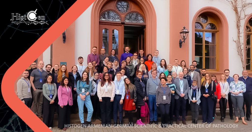
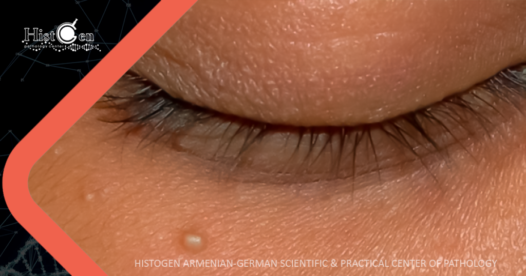
Eng Blog

The 5th Armenian Oncology Annual Congress

The 5th Armenian Oncology Annual Congress was held in Tsaghkadzor, Armenia on February 1-2, 2025, which was also attended by a team of doctors from the Histogen Laboratory. Voskehat Mkrtumyan, a doctor- pathologist of the laboratory, gave a report on “Molecular classification of endometrial carcinoma” at the oncogynecology session. The topic remains crucial nowadays since endometrial carcinoma is the most common gynecological malignancy among women. Author Voskehat Mkrtumyan Voskehat Mkrtumyan The report covered the following topics: the evolution of the classification of endometrial carcinoma, from the earliest anatomic to the most modern molecular, as well as the characteristics, prognostic differences and importance of the molecular types in evaluating the need for adjuvant therapy in carcinoma according to the latest guidelines and updates. Examinations & Services Scanning Scanning Punch biopsy Punch biopsy Pap test Pap test Fine needle aspiration Fine needle aspiration Liquid Based Cytology LBC Liquid Based Cytology LBC Punch biopsy Punch biopsy Histological examination Histological examination Fine needle aspiration Fine needle aspiration Cytological examination Cytological examination Immunohistochemical examination Immunohistochemical examination Pap test Pap test Molecular pathology Molecular pathology As a reminder, the determination of the molecular types presented in the report is carried out at our center (POLE study, immunohistochemical studies). The 5th Armenian Oncology Annual Congress The 5th Armenian Oncology Annual Congress School of Nephropathology School of Nephropathology Ongoing education and the implementation of diagnostic principles following evolving international trends are essential parts … BENIGN LESIONS OF THE EYELID BENIGN LESIONS OF THE EYELID The eyelid (Latin: palpebra) is a thin, movable layer of skin that covers the eye … Following the development of cytology։ internship in Poland Following the development of cytology։ internship in Poland On September 23-27, 2024, the 15th annual Tutorial of Cytology organized by the European Federation …

The 5th Armenian Oncology Annual Congress Read More »

Նեֆրոպաթոլոգիայի դպրոց, Школа нефропатологии

School of Nephropathology

Ongoing education and the implementation of diagnostic principles following evolving international trends are essential parts of our daily practice. Author Armen Mkhitaryan Armen Mkhitaryan On January 25-28, the European Society of Pathologists organized the “School of Nephropathology” in Basel (Switzerland). International experts presented the latest diagnostic principles, the nuances of laboratory processing of nephrobiopsies, and modern methods of interpreting results. Examinations & Services Scanning Scanning Punch biopsy Punch biopsy Pap test Pap test Fine needle aspiration Fine needle aspiration Liquid Based Cytology LBC Liquid Based Cytology LBC Punch biopsy Punch biopsy Histological examination Histological examination Fine needle aspiration Fine needle aspiration Cytological examination Cytological examination Immunohistochemical examination Immunohistochemical examination Pap test Pap test Molecular pathology Molecular pathology To get acquainted with the developments in this narrow field, which requires hypervigilance in terms of diagnosis, a combination of different detection methods at the same time, and versatile knowledge, and to apply them at HistoGen, our center’s chief specialist Armen Mkhitaryan participated in this unique training course held in Basel, and will introduce the acquired experience in Armenia to ensure maximum diagnostic accuracy. School of Nephropathology School of Nephropathology BENIGN LESIONS OF THE EYELID BENIGN LESIONS OF THE EYELID The eyelid (Latin: palpebra) is a thin, movable layer of skin that covers the eye … Following the development of cytology։ internship in Poland Following the development of cytology։ internship in Poland On September 23-27, 2024, the 15th annual Tutorial of Cytology organized by the European Federation … Nevi (moles) Nevi (moles) Nevi (moles) Nevi, mostly known as moles, are benign brown lesions that can be either …

School of Nephropathology Read More »

BENIGN LESIONS OF THE EYELID

The eyelid (Latin: palpebra) is a thin, movable layer of skin that covers the eye from the front, protecting it from external dangerous irritants (ultraviolet rays, dust, etc.). It also keeps the surface of the eyes moist, preventing them from drying out. Author Lilit Meliksetyan Histologically, the following layers of the eyelid are distinguished: skin, subcutaneous connective tissue, muscle layer, tarsal plate, palpebral conjunctiva. In each layer, various lesions can occur: inflammatory, neoplastic, etc. The eyelid lesions can be both non-specific, which also occurring in different parts of the body, and specific, because of the peculiarities of the eyelid tissue. Among of the non-specific lesions, the most common inflammatory disease of the skin of the eyelids is contact dermatitis, which can be caused by various factors: allergic or other physical and chemical irritants. Benign skin non-specific neoplasms on the eyelid may include seborrheic keratosis, simple warts, epidermoid cysts and nevi (moles). Squamous cell papilloma is typical for this area, which is mostly a small benign neoplasm. Xanthelasma is also characteristic in this area, which is a yellowish soft plaque. The occurrence of the latter is mainly associated with a disturbances of fat and cholesterol metabolism. Papilloma Xanthelasma The subcutaneous connective tissue of the eyelid and the tarsal plate contain the largest number of sebaceous glands – the Zeiss and Meibomian glands, which is why various lesions of the sebaceous glands often develop on the eyelid: inflammation (hordeolum), hyperplasia, adenoma. Hordeolum Sebaceous gland hyperplasia Sebaceous gland adenoma As in different areas of the skin, there are sweat glands here, some of which are modified and are called Moll’s ciliary glands. Hydrocystomas, benign cystic formations, can occur due to blockage of sweat glands for various reasons. The latter can be eccrine and apocrine, which differ in size, location and quantity. Hydrocystomas create mainly a cosmetic defect, but to confirm this diagnosis, a histological examination is necessary to exclude a malignant lesion of this area. Apocrine hydrocystoma Eccrine hydrocystoma In the subcutaneous tissue of the eyelid, there are hair follicles from which the eyelashes originate, which also perform a protective function. Therefore, various formations originating from hair follicles can occur here: trichoadenoma, trichofolliculoma, trichilemoma, pilomatricoma, etc. Some of them are prone to malignancy. Malignant formations on the eyelid are rare, but it is important to notice any changes in this area in time and consult a doctor to prescribe appropriate treatment and avoid further complications. Following the development of cytology։ internship in Poland Following the development of cytology։ internship in Poland Nevi (moles) Nevi (moles) AST ISO 9001-2016 Quality assurance is our motto AST ISO 9001-2016 Quality assurance is our motto ASNO Neuro-Oncology 2024 Conference in Singapore: Insights into Neuropathology ASNO Neuro-Oncology 2024 Conference in Singapore: Insights into Neuropathology

BENIGN LESIONS OF THE EYELID Read More »

Following the development of cytology։ internship in Poland

On September 23-27, 2024, the 15th annual Tutorial of Cytology organized by the European Federation of Cytology Societies (EFCS) was held in Poland, in which the pathologist of our center, Tamara Harutyunyan took part. The tutorial was based on on-site microscopy workshops and lectures covering the most important topics in cytopathology, while additional virtual presentations were sent in advance to the participants and discussed live at the end of each practical workshop. Author Tamara Harutyunyan Tamara Harutyunyan The EFCS Annual Tutorial gives cytopathologists and cytotechicians opportunity to participate in an event of high educative and practical value carried out by renowned European tutors. During the event, lectures on cytology of gynecological and non-gynecological (thyroid, breast, salivary gland, fluids accumulating in body cavities, respiratory system, urine, pancreas, lymph nodes) pathologies were presented, after which all the participants had the opportunity to participate in practical seminars, evaluate and discuss both frequent and extremely rare clinical cases, shared by the professors from their experience and from the archives available in the laboratories. After the practical courses, the virtual slides were discussed, and exhaustive answers were given to all the questions. New connections were established, experience was exchanged, and our specialist will apply the acquired knowledge in her daily work to contribute to our center and develop cytology in our country. Nevi (moles) Nevi (moles) The breast biopsy The breast biopsy Following the development of cytology։ internship in Poland Following the development of cytology։ internship in Poland ASNO Neuro-Oncology 2024 Conference in Singapore: Insights into Neuropathology ASNO Neuro-Oncology 2024 Conference in Singapore: Insights into Neuropathology

Following the development of cytology։ internship in Poland Read More »

Nevi (moles)

Nevi (moles) Nevi, mostly known as moles, are benign brown lesions that can be either elevated from the surface of the skin or flat. The cause of the lesion is the nodular proliferation of melanocytes, which synthesize melanin, the pigment responsible for the brown color of the mole. Nevi can be congenital or acquired and are usually not dangerous. However, one must be cautious of changes in certain features, such as color (moles can darken or lighten), size (increase by 2mm or more in a year), asymmetrical and uneven borders, thickening, a feeling of burning, tension, tingling, redness of the skin around the mole, or bleeding of the lesion. If you notice any of the changes listed above, it is important to visit an oncologist, oncodermatologist, or dermatologist. In some cases, changes may be linked to factors such as sun exposure or hormonal changes, which are not necessarily harmful. However, excessive sun exposure can lead to aggressive tumors known as melanomas. Other risk factors for melanomas include UV radiation, chronic skin irritation, exposure to radioactive substances, poor diet, alcohol consumption, smoking, hormonal changes, genetics, light skin (which lacks melanin, affecting skin protection), a high number of moles (more than 50), and immunodeficiency. The presence of one factor does not usually lead to melanomas, but rather a combination of these factors. Melanomas can vary in shape, color, or size and may be symmetrical or asymmetrical. Melanomas typically lack hair, so sudden hair loss from a mole and a halt in growth may be concerning. An increase in pigment is often observed around the lesion. Melanomas often metastasize to lymphatic and blood vessels. The most important prognostic factor is the depth of invasion; the size of the lesion does not affect the prognosis. These lesions typically metastasize to the bones, liver, lungs, and brain, affecting the prognosis. The final diagnosis is made based on the patient’s medical history, dermatoscopic examination, and an incisional or excisional biopsy if necessary, which may be followed by molecular (BRAF mutation) or radiological tests. It is important to avoid self-treatment. Additionally, it is crucial to remember that removing benign nevi does not affect the risk of malignant transformation, and malignant melanomas must be removed. Early detection of melanomas is essential for a better prognosis. AST ISO 9001-2016 Quality assurance is our motto AST ISO 9001-2016 Quality assurance is our motto ASNO Neuro-Oncology 2024 Conference in Singapore: Insights into Neuropathology ASNO Neuro-Oncology 2024 Conference in Singapore: Insights into Neuropathology The breast biopsy The breast biopsy

Nevi (moles) Read More »

AST ISO 9001-2016 Quality assurance is our motto

  AST ISO 9001-2016 Quality assurance is our motto </h1 > Conducting morphological studies following international standards is our priority. “Histogen” Scientific-practical Center of Pathology has once again proven its compliance with the requirements of AST ISO 9001-2016 “Quality Management Systems” and received a certificate from the “National Body for Standards and Metrology ” which guarantees accurate diagnostics ASNO Neuro-Oncology 2024 Conference in Singapore: Insights into Neuropathology ASNO Neuro-Oncology 2024 Conference in Singapore: Insights into Neuropathology The breast biopsy The breast biopsy

AST ISO 9001-2016 Quality assurance is our motto Read More »

ASNO Neuro-Oncology 2024 Conference in Singapore: Insights into Neuropathology

  ASNO Neuro-Oncology 2024 Conference in Singapore: Insights into Neuropathology </h1 > From August 14-18, 2024, the 19th Asian Society for Neuro-Oncology (ASNO) annual meeting was held in Singapore, bringing together top specialists from around the world to discuss the latest research in neuro-oncology. Author Harutyun Papoyan Pathologist From HistoGen lab, we had a pathologist Harutyun Papoyan representing us at this prestigious event, providing an excellent platform for sharing knowledge on the diagnosis and management of brain tumors. One of the most impactful sessions was a full day dedicated to neuropathology, where leading experts presented in-depth case studies on gliomas and meningiomas. These presentations highlighted the diagnostic complexities of these tumors, focusing on differential diagnosis through molecular profiling techniques. Gliomas, often challenging to diagnose, were the subject of several detailed case presentations. Speakers presented how novel genetic markers are aiding in distinguishing these tumors from other central nervous system lesions. For meningiomas, a range of clinical cases was shared, covering various grades and the diagnostic challenges they pose, particularly in distinguishing benign forms from more aggressive variants. Our lab’s pathologist had the opportunity to meet with prominent neuropathologists from Japan, India, Hong Kong, and Singapore. These interactions fostered discussions on clinical cases, diagnostics, and emerging trends in the field, paving the way for potential research collaborations. The knowledge and experience gained at the ASNO 2024 conference have enriched our lab’s approach to neuro-oncology research, particularly in understanding the subtleties of diagnosing gliomas and meningiomas. We look forward to incorporating these insights into our ongoing work and building on the connections made during the event. We would like to extend our heartfelt thanks to Ibrahim Qaddoumi, MD, MS and St. Jude hospital for the opportunity to participate in this incredible event and contribute to the future of neuropathology. The breast biopsy The breast biopsy

ASNO Neuro-Oncology 2024 Conference in Singapore: Insights into Neuropathology Read More »

Կրծքագեղձի բիոպսիա, Биопсия молочной железы

The breast biopsy

  The breast biopsy </h1 > The breast biopsy is a medical manipulation procedure where a small sample is taken from breast tissue, which is then examined under the microscope by a pathologist. author Voskehat Mkrtumyan Pathologist The biopsy is performed when a lesion is suspected or detected either on sonography or mammography and it is necessary to find out whether the lesion is benign or malignant or else there might be an inflammatory process. After the microscopic examination of the sample the pathologist can tell what kind of lesion it is. If it is a cancer, then a further immunohistochemical examination is needed to distinguish the subtype of the tumor. During that examination the amount of hormonal receptors or Her2neu status and Ki67 cell division activity in tumor cells are evaluated. Molecular subtype is needed for further neoadjuvant, hormonal or targeted chemotherapies, sometimes also the combination of all three. Sometimes Her2neu status is evaluated as equivocal. In those cases another examination which is called FISH testing is needed, which evaluates if mutation is present in the Her2neu gene. Positive Her2neu status allows the clinician to perform neoadjuvant hormonal chemotherapy with a drug called Herceptin. It is important to mention that after biopsy benign lesions do not become malignant and malignant ones cannot become more aggressive and the biopsy is necessary and irreplaceable if there are medical indications. After the procedure hematomas, bleeding or pain can follow. Patients often wonder if the process is painful. The ones who have gone through the procedure say that what they felt was more of discomfort rather than pain, although it is a subjective sensation and can be varied. Moreover, it is essential to mention that due to modern medicine achievements, breast cancer can be treated in most cases if detected in time. For a probable early detection monthly self-observation is needed, however, one should consider that not all the lesions can be palpated, besides, preventive measures should be taken: sonography (if the patient is 30-40 years old) or mammography (if the patient is 40 years old or above). Կրծքագեղձի բիոպսիա Կրծքագեղձի բիոպսիա Նևուսներ (խալեր) Նևուսներ (խալեր) Մոլեկուլային պաթոլոգիա Մոլեկուլային պաթոլոգիա Պապ թեստ Պապ թեստ

The breast biopsy Read More »